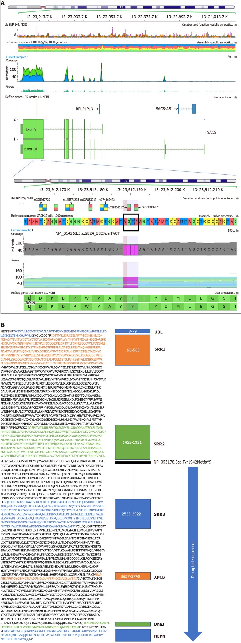
Figure 4.
SACS gene location and N- to C-terminal of the sacsin protein. A: Exome sequencing summary layout depicting the SACS gene location on chromosome 13 at the top and the exons coverage in green and blue. The location of the mutation on exon 10 is represented by a vertical black line. The deletion TACT mutation in the zoomed view is shown in magenta, and the reference sequence marked with a rectangle; B: N- to C-terminal of the sacsin protein, depicting a ubiquitin-like domain that binds to the proteosome, three sacsin repeat regions with known Hsp90-like chaperone function, an xeroderma pigmentosum complementation group C-binding domain that binds to the Ube3A ubiquitin protein ligase, a DnaJ domain that binds Hsp70, and a higher eukaryotes and prokaryotes nucleotide-binding domain that facilitates sacsin protein dimerization. The horizontal black l line depicts the start of the mutation, which causes loss of function of the protein due to the loss of all amino acids downstream of the line. The blue arrow shows the disrupted protein sequences and domain of sacsin protein. Each domain is demarcated by a specific sequence highlighted by the corresponding color and amino acid number given that methionine is the first amino acid at the N-terminus.

