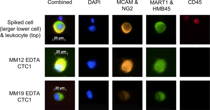
Fig. 2.

Images of spiked SK‐MEL28 cells in HD blood and CTCs identified in the MM patients using a multicolor immunofluorescence approach. After the isolation with the ClearCell system, the immunofluorescence staining was performed using a combination of four different MM markers (MCAM and NG2 in the PE and MART1 and HMB45 with Alexa 488). As an exclusion marker, CD45 (APC) was used. For nuclear staining, DAPI was used (blue channel). CTCs were positive for the melanoma markers and negative for CD45, while leukocytes were only positive for CD45 and negative for the melanoma markers.
