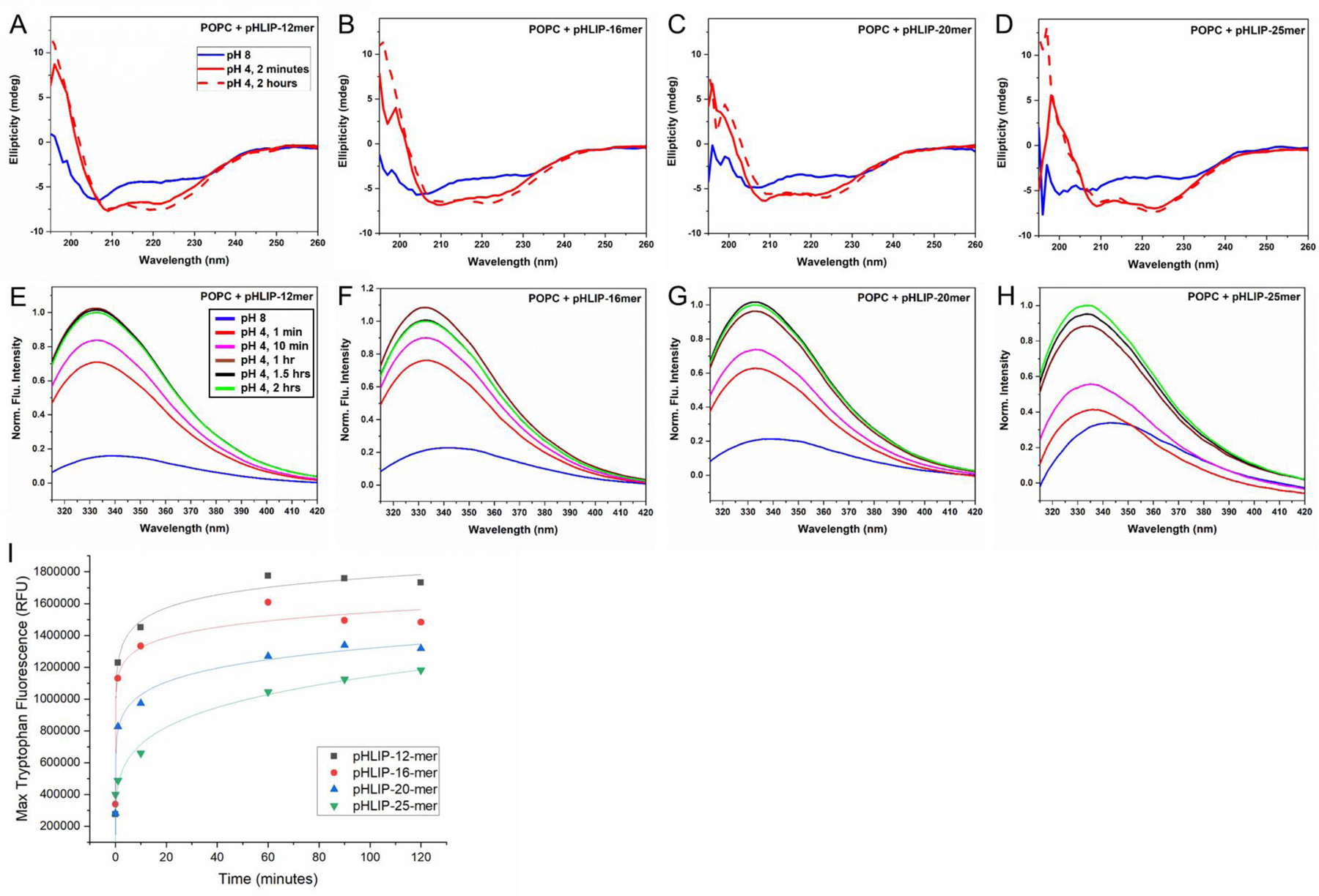
Figure 2: Interactions of pHLIP-PNA constructs with lipid bilayers.

CD spectroscopy (A-D) and tryptophan fluorescence measurements (E-H) at various time points before and after reducing pH from 8 to 4 suggest that pHLIP-PNA constructs incubated with POPC vesicles undergo pH-triggered alpha-helix formation and transmembrane insertion, respectively. Maximum tryptophan fluorescence over time measurements (I) reveal differential kinetics of insertion for various pHLIP-PNA constructs.
