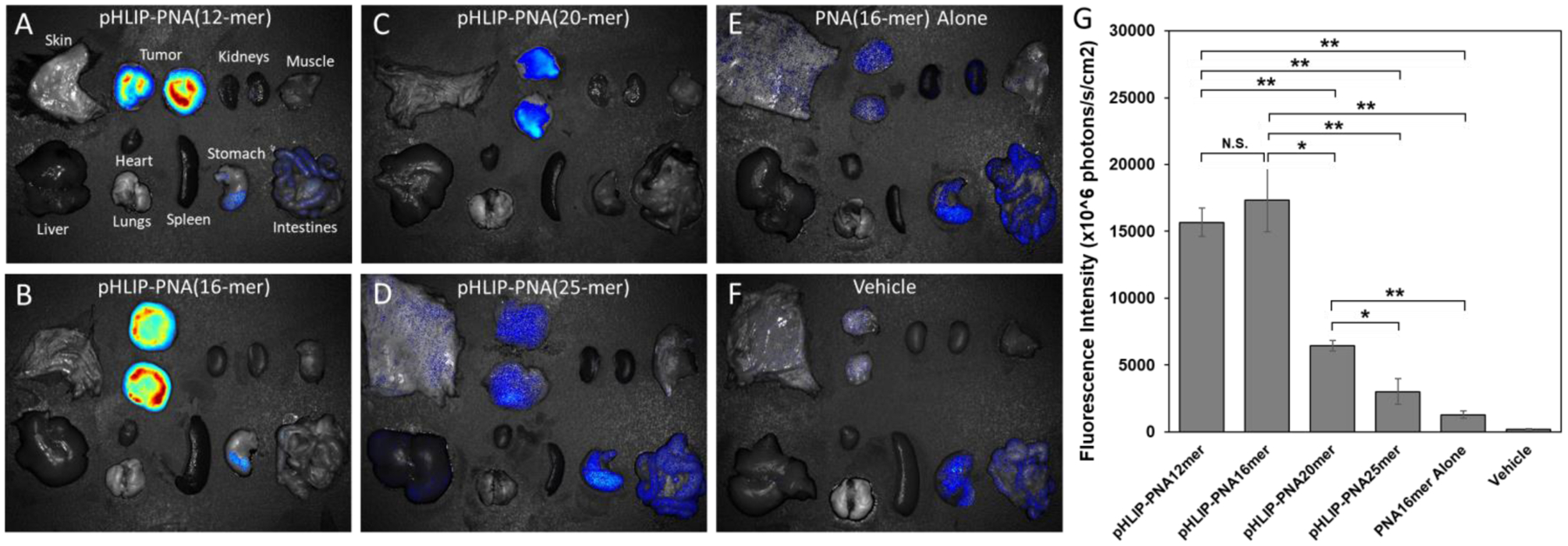
Figure 5: Targeted delivery of PNA cargoes to tumors by pHLIP.

pHLIP-PNA constructs were injected intravenously into melanoma tumor-bearing mice. 24 hours later, the mice were sacrificed and their tumors and major organs were excised and imaged for TAMRA fluorescence (A-F). Prior to imaging, the tumors were split in half to avoid obstruction of the fluorescence by the overlying skin, and the fluorescence intensities per unit area of the resulting two halves were averaged (G). Data are shown as mean ± S.E. (n = 3); * P < 0.05; ** P < 0.01. Of note, the signal in the stomach and intestines is present in the vehicle sample, indicating that it is tissue autofluorescence.
