Fig. 1.
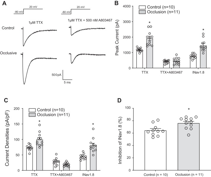
Effects of femoral artery occlusion on TTX-resistant (TTX-R) and voltage-dependent Na+ channel subtype NaV1.8 (NaV1.8) currents in rat dorsal root ganglion (DRG) neurons. All rat DRG neurons were dissociated 72 h after femoral occlusion and recorded in voltage patch mode with −80 mV holding followed by a pulse of 20 mV. One micromolar TTX was used to block TTX-sensitive currents, and 500 nM of A803467 was used to block NaV1.8 currents. A: representative current traces after application of TTX and TTX plus A803467 in DRG neurons of control limb and occluded limb. A greater amplitude of currents was observed in DRG neurons of the occluded limb. B: averaged data showing that femoral artery occlusion amplified current amplitude after TTX application. The data also show that the currents were largely decreased after application of TTX and A803467. NaV1.8 currents were assessed as (peak current after TTX − peak currents after both TTX and A803467). was also amplified by femoral artery occlusion. *P < 0.001 between control and occlusion for TTX-R and NaV1.8 current in DRG neurons. C: femoral artery occlusion also increased current densities in DRG neurons compared with the control group. *P = 0.002 between control and occlusion after TTX; *P < 0.001 between control and occlusion for . D: %inhibition of by A803467 was greater in DRG neurons of occluded limbs than that in DRG neurons of control limbs. *P = 0.010 between control and occlusion. Individual data points are also shown; n = 10 DRG neurons in the control group, and n = 11 in the occlusion group.

