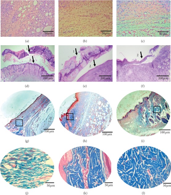
Figure 6.

Inflammatory infiltrate and fibroblasts on the 9th day after lesion treatment of the groups. (a) BFt, (b) Fibrinase®, and (c) FtEHJ; the FtEHJ group showed less inflammation cells in tissue. (d) BFt, (e) Fibrinase®, and (f) FtEHJ;Hematoxylin and Eosin staining, using an optical microscope, 400x magnification. (1) Granulation tissue; (2) reepithelialization. The granulation tissue regressed in the FtEHJ group, while in the other groups, 100x magnification was still used. (g) BFt, (h) Fibrinase®, and (i) FtEHJ; Masson's Trichrome staining, using an optical microscope, 200x magnification. Marked area with squares is presented in higher magnification. When enlarging the images, it is noted that the (j) BFt group has collagen fibers arranged in parallel and with less intense coloring, characteristic of less dense fibers; the (k) Fibrinase® group has dense and disorganized collagen fibers, but the tissue is still moderately vascularized; and in the (l) FtEHJ group, this vascularization was already discreet, and the collagen fibers had the same characteristics found in the Fibrinase group® (400x magnification). Bft, basis of the topical formulation; FtEHJ, topical formulation of the hydroalcoholic extract of J. decurrens Cham.
