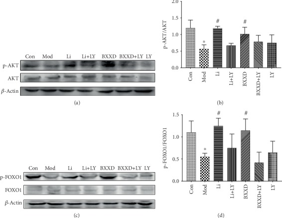
Figure 6.

Effects of BXXD on the expression of AKT, phosphorylated AKT, FOXO1, and phosphorylated FOXO1 in MIN6 cells treated with THBP. (a, b) Western blot and quantitative measurement of AKT and phosphorylated AKT in MIN6 cells. Lane loading was normalized by reblotting with β-actin. The levels were expressed as p-AKT/AKT and normalized relative to the control group. (c, d) Western blot and quantitative measurement of FOXO1 and phosphorylated FOXO1 in MIN6 cells. Lane loading was normalized by reblotting with β-actin. The levels were expressed as p-AKT/AKT and normalized relative to the control group. MIN6 cells were incubated with DMEM (Con), DMEM plus t-BHP (Mod), DMEM, t-BHP plus liraglutide (Li), DMEM, t-BHP, liraglutide plus LY294002 (Li+LY), DMEM, t-BHP plus BXXD 0.5 mg/ml (BXXD), and DMEM, t-BHP, BXXD 0.5 mg/ml plus LY294002 (BXXD+LY). ∗P < 0.05 versus control group; #P < 0.05 versus model group.
