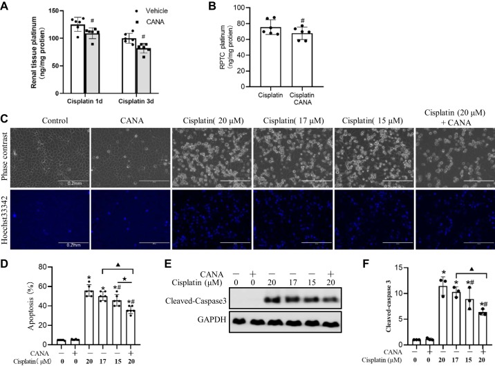
Fig. 5.
Partial inhibitory effect of canagliflozin (CANA) on cisplatin uptake by kidney tissues and cells. A: platinum quantification in mouse kidneys. C57BL/6 mice with or without CANA treatment were euthanized at 1 day (1d) or 3 days (3d) after cisplatin treatment. Data are presented as means ± SD; n = 6. #P < 0.05 vs.the cisplatin only-treated group at the same time point. B: platinum quantification in rat kidney proximal tubular cells (RPTCs). RPTCs were incubated with 20 μM cisplatin in the absence or presence of CANA for 10 h. Values are means ± SD; n = 6. #P < 0.05 vs. the cisplatin-treated group. C: representative cell and nuclear images after Hoechst 33342 staining. Scale bar = 0.2 mm. D: percentage of apoptosis in RPTCs. E: representative immunoblots of cleaved caspase-3. GAPDH was used as an internal loading control. F: densitometric analysis of cleaved caspase 3 normalized to GAPDH. *P < 0.05 vs. the control group; #P < 0.05 vs. the 20 μM cisplatin-treated group; ▲P < 0.05 vs. the 17 μM cisplatin-treated group; ★P < 0.05 vs. the 15 μM cisplatin-treated group.

