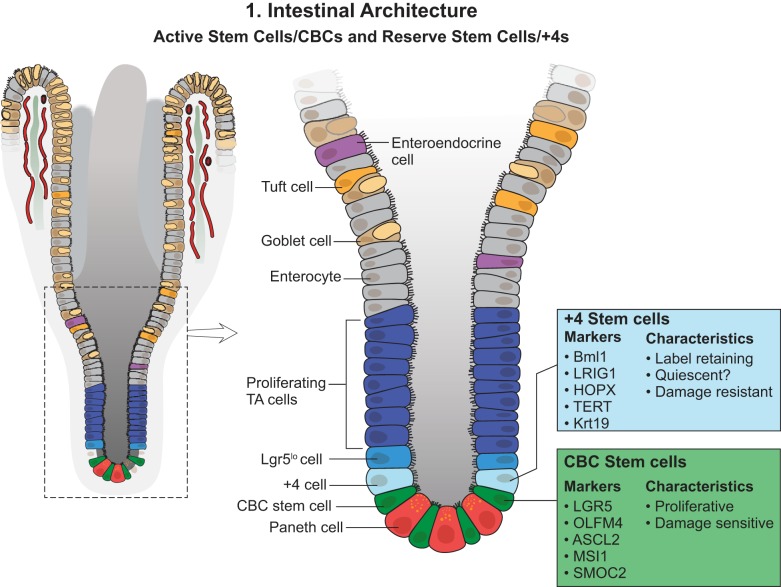
Fig. 1.
Small intestinal structure. The small intestine consists of monolayer epithelial cells covering invaginated gland structures (crypts) and finger-like protrusions (villi). Intestinal stem cells (ISCs) reside in the bottom of the crypts, composed of crypt base columnar cells (CBCs) (active stem cells) and +4 stem cells (reserve stem cells), which could be distinguished by their certain markers but mainly are based on cell cycle status and functional roles. Absorptive and secretory progenitor cells are located above the ISC zone, and more differentiated cells are at the villi. TA, transit amplifying; Lgr5, leucine-rich repeat-containing G protein-coupled receptor 5; BMI1, polycomb group RING finger protein-4; LRIG1, leucine-rich repeats and immunoglobulin-like domains protein-1; HOPX, homeodomain-only protein homeobox; TERT, telomerase reverse transcriptase; Krt19, keratin, type I cytoskeletal 19; OLFM4, olfactomedin 4; ASCL2, achaete-scute complex homolog 2; MSI1, Musashi RNA-binding protein-1; SMOC2, SPARC-related modular calcium-binding protein-2.

