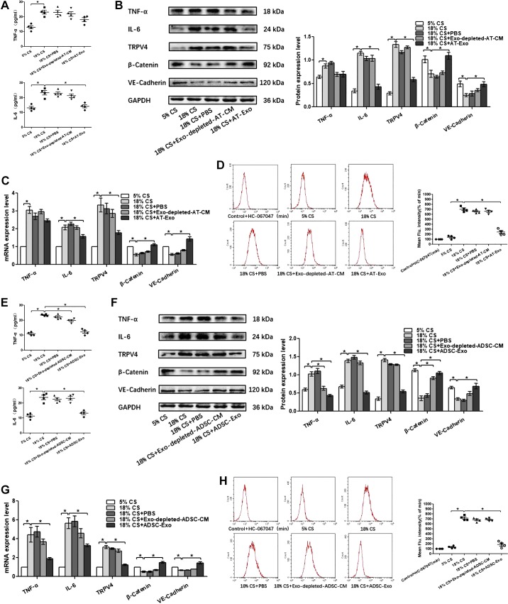
Fig. 8.
Adipose tissue exosome (AT-Exo) and adipose-derived stem cell exosome (ADSC-Exo) promote pulmonary microvascular endothelial cell (PMVEC) barrier function and suppress inflammation after 18% cyclic stretching (CS) through inhibiting transient receptor potential vanilloid 4 (TRPV4)/Ca2+ signaling in vitro. A and E: proinflammatory cytokines TNF-α and IL-6 in cell culture supernatant. B and F: Western blot (WB) analysis shows the protein expression of TNF-α, IL-6, TRPV4, β-catenin, and VE-cadherin, and the relative values are normalized to GAPDH. C and G: quantitative real-time PCR (qRT-PCR) analysis shows the gene expression of TNF-α, IL-6, TRPV4, β-catenin, and VE-cadherin normalized to the gene expression of GAPDH. D and H: intracellular calcium ions are detected by flow cytometry (FCM); n = 4 cultures from each group assayed in triplicate. The results are expressed as means ± SE. *P < 0.05. ADSC-CM, ADSC conditioned media.

