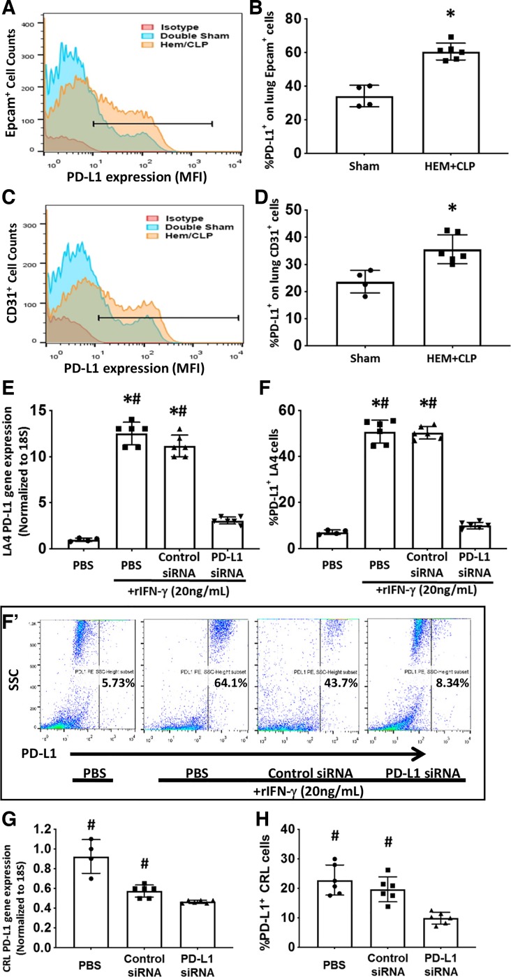
Fig. 2.
Hemorrhagic shock followed by cecal ligation and puncture (Hem-CLP) enhanced PD-L1 expression on lung epithelial and endothelial cells in vivo and PD-L1 siRNA suppresses PD-L1 gene/protein expression in CRL-2167 (CRL) and stimulated LA-4 cells in vitro. PD-L1 protein expression on Epcam+ or CD31+ lung cells was determined by flow cytometry. Typical PD-L1 expression/mean fluorescent intensity (MFI) histogram for those cells positive for either the epithelial cell (A), Epcam+, or the endothelial cell (C), CD31+, markers. The horizontal “line” in those histograms identifies cells determined to be positive for both PD-L1+Epcam+ or PD-L1+CD31+ (from which the %PD-L1+Epcam+ or %PD-L1+CD31+ was established) based on “isotype” antibody-chromophore control. The frequency of PD-L1+Epcam+ or PD-L1+CD31+ was significantly increased on lung epithelial (B) and endothelial (D) cells from Hem-CLP compared with sham-operated mice (means ± SE; n = 4–6 mice per group; *P < 0.05 vs. sham group, rank-sum test). To verify the efficacy of PD-L1 siRNA, the mouse lung epithelial cell line LA4 [E–F (F’)] and the endothelial cell line CRL (G and H) were used. PD-L1 mRNA and protein were determined following siRNA transfection. Subsequently, the expression of PD-L1 gene (E) and protein (F) [representative dot plots of LA4 cell side-scatter (SSC) profile vs. PD-L1 staining intensity for each condition summarized for multiple experiments is shown in F’] of LA4 cells were increased after rIFN-γ stimulation and reduced with anti-PD-L1 siRNA. Endothelial cell line CRL, which constitutively expresses PD-L1, when treated with anti-PD-L1, but not Control, siRNA treatment reduced PD-L1 gene (G) and protein (H) expression (means ± SE; n = 4–6 individual experiments/group; *P < 0.05 vs. PBS alone group; #P < 0.05 vs. PD-L1 siRNA-treated group, by one-way ANOVA and a Student–Newman–Keuls test).

