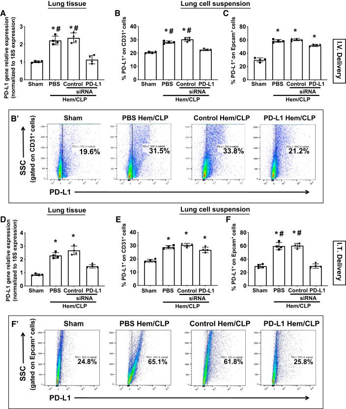
Fig. 3.
Differential effects of intravenous (I.V.) versus intratracheal (I.T.) delivered PD-L1 siRNA on suppressing PD-L1 expression in the lungs of mice with indirect acute lung injury (iALI). The lungs were collected 24 h after hemorrhagic shock followed by cecal ligation and puncture (Hem-CLP) or sham operation. Lung tissue homogenate lysates were used for PD-L1 gene expression. PD-L1 gene expression was upregulated in all iALI mice compared with sham mice and was selectively suppressed by anti-PD-L1 siRNA when delivered either intravenously (A) or intratracheally (D). Enzymatic digested lung single-cell suspensions were examined for PD-L1 protein expression by flow cytometry. Intravenous delivery of siRNA suppressed PD-L1 expression by CD31+ lung endothelial cells (B and B′: a representative dot plot of CD31+ cells), but not by Epcam+ lung epithelial cells of Hem-CLP mice (C). Alternatively, intratracheal delivery of siRNA inhibited the PD-L1 expression on Epcam+ lung epithelial cells (F and F′: a representative dot plot of Epcam+ cells), but not CD31+ lung endothelial cells (E). Values are means ± SE; n = 4–6 mice per group; *P < 0.05 vs. Sham; #P < 0.05 vs. PD-L1 siRNA-treated Hem-CLP group, by one-way ANOVA and a Student–Newman–Keuls test.

