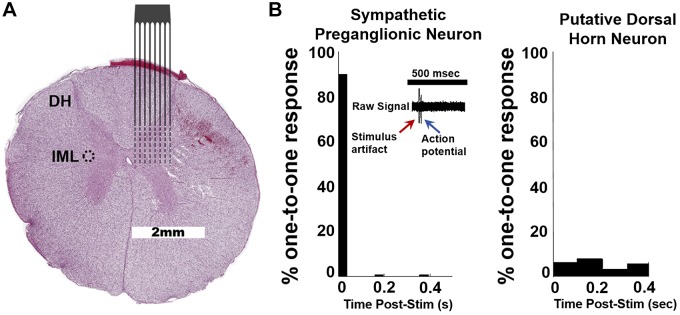
Fig. 1.
A: representative porcine spinal cord (T2; hematoxylin and eosin staining) with overlay of approximate location of recording electrode array. Intermediolateral nucleus (IML; circled). B: T2 paravertebral stimulation elicits spike firing in IML to identify sympathetic preganglionic neurons (SPNs). Stimulation should elicit a one-to-one response in SPNs (left) vs. other, putative dorsal horn (DH) neurons (1,703 neurons; 155 ± 48 per pig; n = 10). SPNs were identified as those that exhibited a one-to-one response to T2 paravertebral stimulation within <0.05 s at least 60% of the time. SPNs were only identified in 8 of 10 pigs (57 neurons; 6 ± 2 per pig; n = 8).

