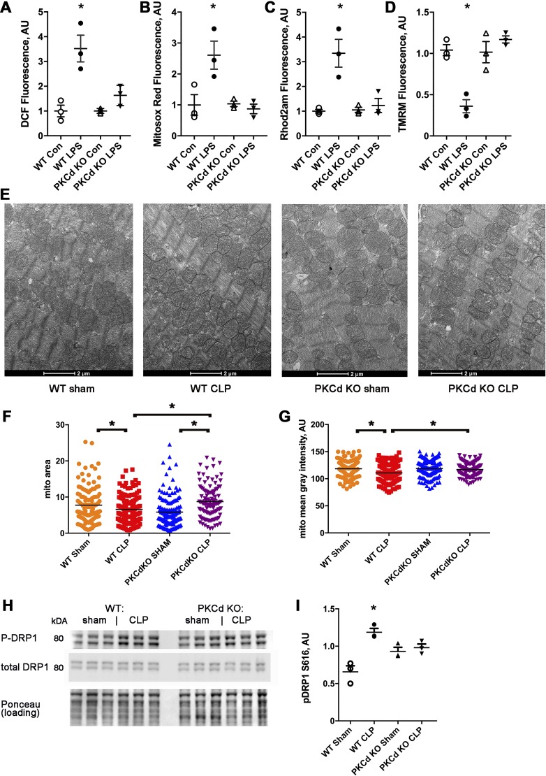
Fig. 5.
PKCδ knockout (KO) cardiomyocytes are protected from mitochondrial dysfunction induced by lipopolysaccharide (LPS) and mitochondrial morphologic changes after cecal ligation and puncture surgery (CLP). A: isolated cardiomyocytes, 2',7'-dichlorofluorescein (DCF) fluorescence minus background, n = 3 wells, 2,000 cells/well, bars represent means + SE. AU, arbitrary units. The means of the groups are different by ANOVA, *significantly different from control (Con) by post hoc test. B: cardiomyocytes, MitoSOX red readout. C: cardiomyocytes, Rhod 2-AM signal, and mitochondrial calcium. D: cardiomyocytes, tetramethylrhodamine methyl (TMRM) signal to indicate mitochondrial inner membrane potential. E: representative electron microscopy from ventricular samples of wild-type (WT) sham, WT CLP, and PKCδ KO. F: mean mitochondrial area. G: mean mitochondrial pixel density. B and C: N = 100–140 mitochondrial from each group, n = 4 hearts each group, with 3 images from each heart sample. The means of the groups are different by ANOVA, *significantly different by post hoc test. H: representative Western blots of phospho-dynamin-related protein 1 (Drp1) and total Drp1. I: quantification of phospho-Drp1 normalized to total Drp1. The means of the groups are different by ANOVA, *significantly different by post hoc test.

