Fig. 1.

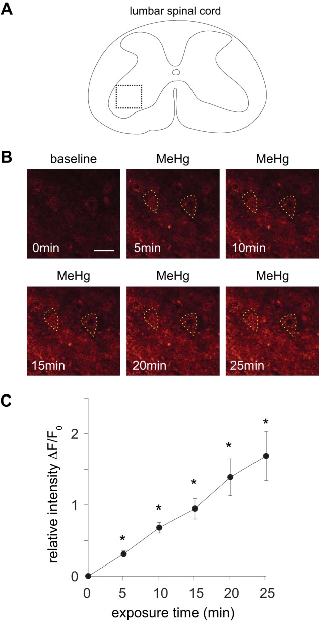
Methylmercury (MeHg) induced an increase in fluo 4 fluorescence in lumbar spinal motor neurons (MNs). A: illustration of a lumbar spinal cord slice imaged with fluo 4-AM in the presence of 2 mM Ca2+. Dashed inset reflects the region of the spinal cord ventral horn shown in B. B: microfluorometric images (scale bar, 50 μm) of lumbar spinal cord motor region labeled with fluo 4-AM before (0 min) and after exposure to 20 μM MeHg (5–25 min). Individual spinal MNs are outlined with dashed line. C: relative fluorescence intensity (ΔF/F0) is shown vs. exposure time to 20 μM MeHg across the imaged spinal cord slices (n = 5) for the population of identified spinal MNs (n = 15). There was a significant increase (*P = 4.1e-6) in ΔF/F0 across the sampled MNs of the ventral spinal cord after MeHg exposure (t > 10 min).
