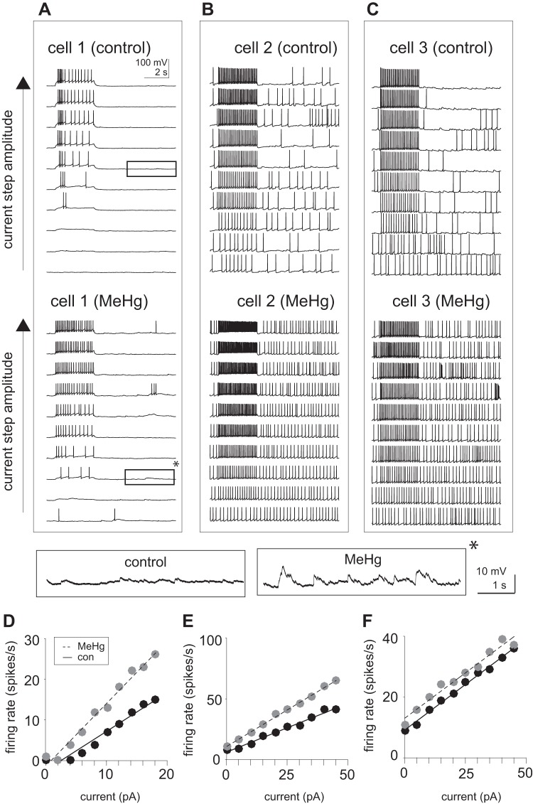
Fig. 2.
Methylmercury (MeHg) increased spontaneous and evoked action potential generation in lumbar spinal motor neurons (MNs). Current injections were used in current-clamp mode to estimate firing rate vs. current (f-I) relationships. A–C: representative voltage recordings (10 s) of spinal MNs in response to increasing current-step (3 s) amplitudes before and after MeHg exposure. MeHg induced both an increase in the firing rate to the current step as well as the spontaneous spiking (occurring outside of the time of the current pulse). Insets (bottom) indicate close-up views of membrane potential during spontaneous (nonstimulated) period. The amplitude of fluctuations in voltage are significantly enhanced in the presence of MeHg (asterisk). D and E: MeHg induced a significant increase in the responsiveness to current injections. While some cells (cell 1 and cell 2; D and E, respectively) showed an increase in f-I slope, others (cell 3; F) showed an upward shift in f-I without a change in slope. Estimates of action potential rate are shown as black circles for control and gray circles for MeHg-exposed conditions. Solid and dashed lines represent linear regression fits for control and MeHg responses, respectively.

