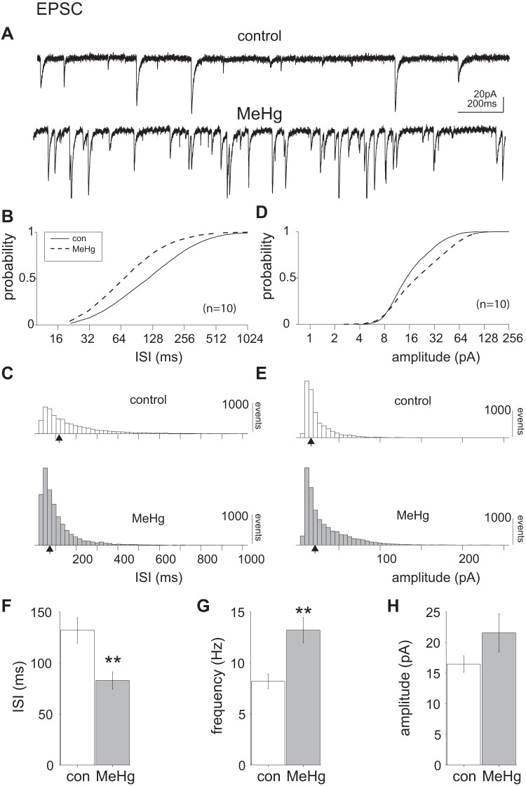
Fig. 4.
Methylmercury (MeHg) significantly increased excitatory postsynaptic currents (EPSCs) in lumbar spinal motor neurons (MNs). A: representative voltage-clamp recordings of a single lumbar spinal MN before and after 20 μM MeHg exposure. After 15 min of MeHg exposure, there was a noticeable increase in spontaneous EPSC (sEPSC) frequency. B and C: comparison of control and MeHg-exposed conditions (solid and dashed lines, respectively) across the population of sampled neurons (n = 10 neurons). The intersynaptic interval (ISI) cumulative distribution is significantly shifted to the left (P = 2e-209, Kolmogorov–Smirnov test). D and E: there was also a significant rightward shift in the cumulative amplitude distribution (P = 1.4e-104, Kolmogorov–Smirnov test). F–H: across the population there was also a significant (**P = 0.0091, Wilcoxon rank sum test) decrease in the mean ISI (132 ± 12 and 82 ± 8 ms for control and MeHg-treated neurons, respectively), corresponding to an increase in sEPSC mean frequency from 7.5 ± 0.7 Hz (control) to 12.1 ± 1.2 Hz (MeHg). The mean population amplitudes (16.6 ± 1.3 and 21.6 ± 3 pA, control and MeHg, respectively) were not significantly different, despite the trend. Open and shaded bars represent control and MeHg conditions, respectively. con, Control. Black arrows indicate the geometric mean of the distribution.

