
Figure 8.
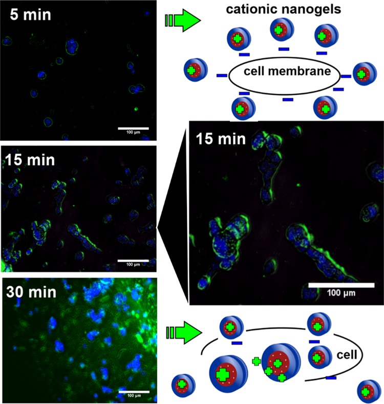
Cell-internalization images using fluorescence microscopy of fluorescent PDEAEM-core-PEG-shell nanogels (100 μg/mL for 5, 15, and 30 min of incubation); cell nuclei were counterstained with Hoechst 33258 in blue, and fluorescent nanogels (NF1) were visualized in green.