
FIGURE 1.
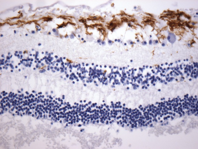
Sample immunohistochemical stains at 40x show good antibody penetrance for GFAP in retinal tissue, with positive staining of Müller glia.
Official websites use .gov
A
.gov website belongs to an official
government organization in the United States.
Secure .gov websites use HTTPS
A lock (
) or https:// means you've safely
connected to the .gov website. Share sensitive
information only on official, secure websites.
Sample immunohistochemical stains at 40x show good antibody penetrance for GFAP in retinal tissue, with positive staining of Müller glia.