
Figure 2.
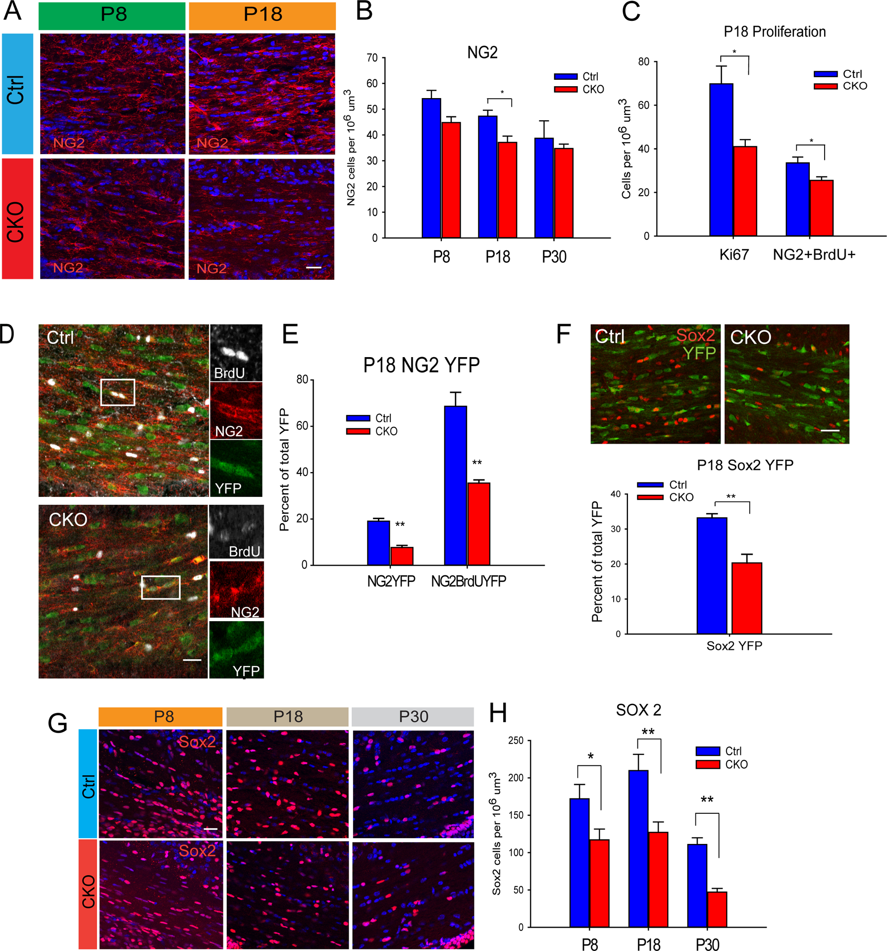
Sox17 regulates OPC expansion. (A–B) Immunohistochemical analysis of NG2-expressing OPCs in P8 and P18 WM shows little change at P8, but significant decrease at P18. (C) Analysis of proliferation using Ki67 and BrdU reveals decreased Ki67 and BrdU labeling in WM at P18. N=4. *p < 0.05, and **p < 0.005, Student’s unpaired T test, mean ± SEM. 50mg/kg BrdU was injected i.p. 24 hr prior to sacrifice. (D) Decreased total NG2 and BrdU-labeled OPCs within the CNP-Cre;RosaYFP population at P18 indicates an underlying deficit in proliferation (E). N=3. *p < 0.05, and **p < 0.01, Student’s unpaired T test, mean ± SEM. (F) The number of Sox2-expressing progenitor cells is decreased in P18 CKO WM. N=3. *p < 0.05, and **p < 0.005, Student’s unpaired T test, mean ± SEM (G) The total number of Sox2-expressing cells is decreased in developing WM of the postnatal CKO. N=4 **P<0.005, *P<0.05 Student’s T-test vs Ctrl. Scale bars: A,D,F 20 um, G,50 um.