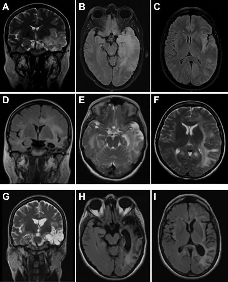
Figure 2.
Brain magnetic resonance imaging (MRI) evolution of the first patient. Initial MRI—coronal T2 imaging (A) and axial fluid-attenuated inversion recovery (FLAIR) imaging (B-C) showing hyperintensity in the left temporal lobe involving the hippocampus and hyperintensity in left temporal and insular lobes, respectively; post-herpes simplex virus encephalitis (HSVE) anti-N-methyl-d-aspartate receptor (anti-NMDAR) encephalitis—coronal FLAIR imaging (D) showing extensive left temporal and insular lobes hyperintensity with dilatation of temporal horn and choroid fissure; axial T2 imaging (E-F) with bilateral hyperintensity in temporal and insular lobes, with predominance in the left side with parietal and frontal extension; follow-up imaging (after immunotherapy and approximately 10 weeks after initiation of acyclovir)—coronal T2 imaging (G) showing left temporal region of increase T2 signal with cystic degeneration and ex-vacuo dilatation of temporal horn; axial FLAIR imaging (H-I) with left temporoparietal gliosis.

