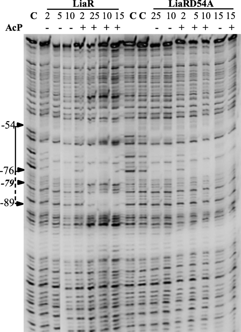
Fig. 7.

LiaR-DNase I footprint analysis of PliaI. The DNA fragment was labeled on the top strand at the 5′-end with [γ-32P]-ATP. The position of the protection site in the gel was determined from the running of the A, T, G and C standard reactions (Additional file 8: Figure S8). Protection of PliaI by LiaR and LiaRD54A was investigated in the absence and presence of acetyl phosphate and at different protein concentrations. The primary binding site is indicated by a solid line, and the secondary binding site is indicated by a dashed line. The hypersensitive site at − 79 is indicated by an arrow
