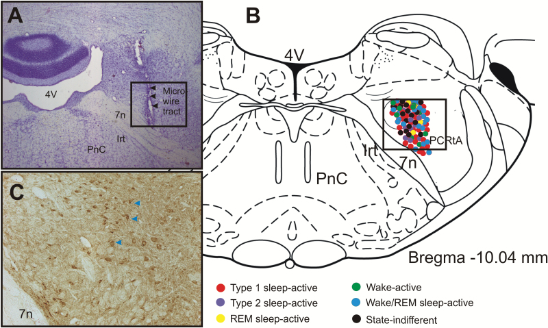
Figure 1.
Site of extracellular recording and anatomical distribution of various sleep-wake neuronal phenotypes. (A). Photomicrograph of a representative coronal section (Nissl stained) through the parafacial zone showing the microwire tracts indicated by arrows. (B) Drawing of a representative coronal section through the parafacial zone [24] showing the anatomical distribution of the extracellularly recorded neurons along with their sleep-wake discharge profiles. Although, the rostrocaudal plane of the recorded neurons varied by 100–250 µm through the parafacial zone, they have been superimposed on a single plane of section for simplicity. (C) Photomicrograph of a representative coronal section through the parafacial zone showing the distribution of GAD67-immunopositive neurons (brown cells, arrows) in a comparable area as marked in figures (A) and (B).  , type 1 sleep-active neurons;
  , type 2 sleep-active neurons;
  , REM sleep-active neurons;
  , wake active neurons;
  , wake/REM sleep-active neurons; and
  , state-indifferent neurons. Note that most of the recorded neurons were localized in the GABA-immunopositive neuronal field. 7n, facial nerve; 4V, fourth ventricle; IRt, Intermediate reticular nucleus; PCRtA, parvicellular reticular nucleus, alpha part; PnC, pontine reticular nucleus.

