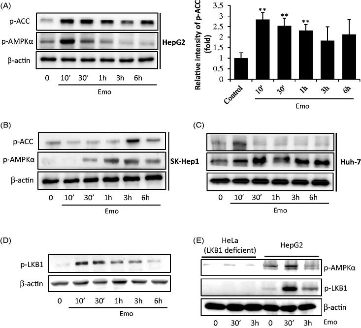
Figure 3.
AMPKα activation by emodin. HepG2 cells were incubated in serum free media for 12 h, and then treated with 30 μM Emo for indicated time periods. Western blotting analysis of p-ACC, p-AMPKα and β-actin and band intensity of p-ACC (**p < 0.01 between vehicle and Emo-treated cells). Western blotting analysis was performed with HepG2 (A), SK-Hep-1 (B) and Huh-7 cells (C). (D) Expression of p-LKB1 in HepG2 cells. (E) Compare of LKB1-deficient Hela cells and HepG2 cells. Emo: emodin.

