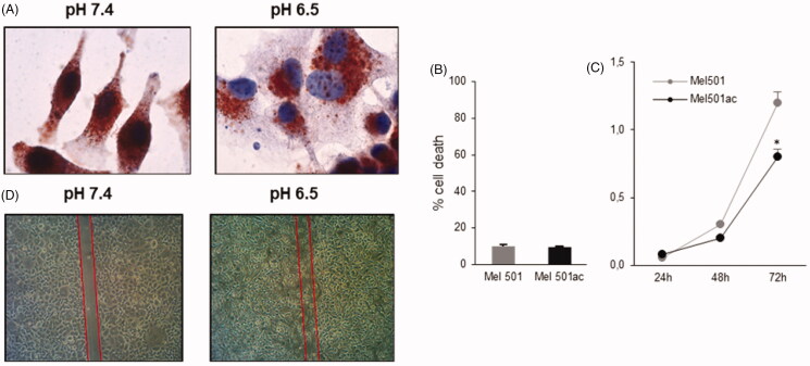
Figure 1.
Analysis of Mel501 vitality and growth properties in buffered (Mel501) and in acidic conditions (Mel501ac). (A) Immunocytochemistry assay. Cells were fixed in cold 70% methanol and stained with mouse monoclonal anti-LAMP-2 using the peroxidase anti-peroxidase method in single staining. Images were acquired at 60× original magnification. (B) Cell viability assay. Cells were stained with 0.05% Trypan Blue for 10 min and cell death was determined by flow cytometry. The percentage of cells incorporating Trypan Blue with respect to total cells counted is reported. (C) Growth curve of Mel501 adapted at low pH. The cell number is reported. Mean values were calculated on 3 replicates and mean ± s.d. is indicated for each sample. *p < 0.05. (D) Wound closure assay. Cells were scraped, and debris washed with PBS. After 18 h, dishes were observed under a phase-contrast microscope and images were acquired, 10× original magnification.

