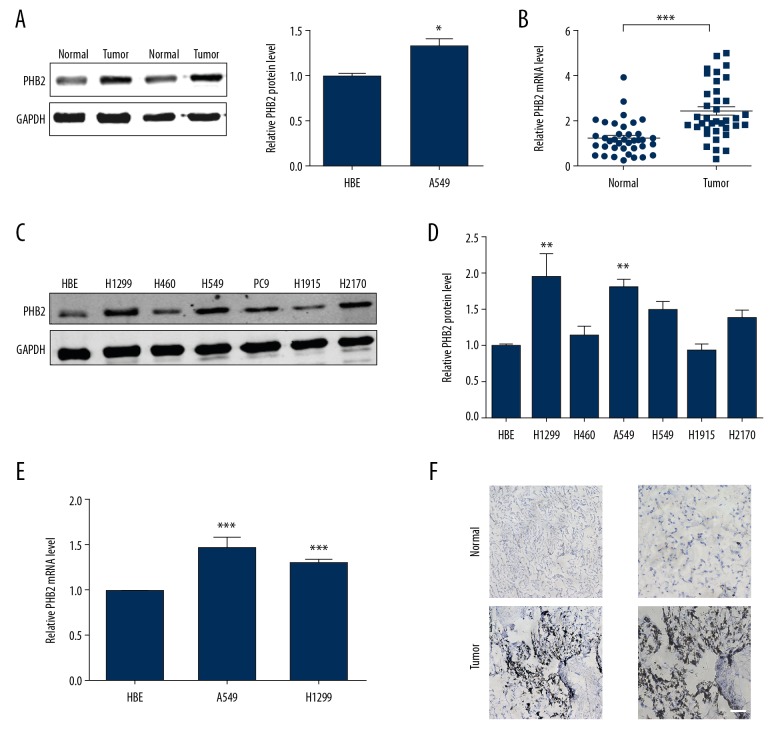
Figure 1.
PHB2 expression in NSCLC. (A) PHB2 expression in human tissues was detected by Western blot (n=5). * P<0.05 vs. Normal. (B) mRNA expression of PHB2 in human tissues were measured by qRT-PCR (n=38). *** P<0.001 vs. normal. (C, D) PHB2 protein expression in H1299, H460, A549, PC9, H1915, H2170, and HBE cells were measured (n=5). *** P<0.01 vs. HBE. (E) The relative quantities of PHB2 mRNA in A549, H1299, and HBE cells were measured (n=5). ** P<0.01 vs. HBE, *** P<0.001 vs. HBE. (F) Expression of PHB2 protein in human NSCLC and paired normal tissues was detected by immunohistochemistry (n=5). Representative pictures are shown. Scale bar indicates 100 μm.

