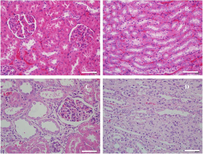
Figure 6. Microscopic histology of rat kidneys in male wild-type rats (A, B) and Uox−/− rats (C, D).
Nephrocytes in wild-type rats were piled tightly (A, B); and there were no swelling signs in glomeruli (A) and tubules (B). Meanwhile in Uox−/− rats, cells in the glomeruli were slightly swollen and their capsular spaces were slightly dilated (C), the tubular walls became thin, and their spaces were also slightly dilated (D). Sporadic interstitial fibrosis and inflammatory cell infiltration can also be seen in the kidneys of Uox−/− rats (C, D). Bar = 50 µm.

