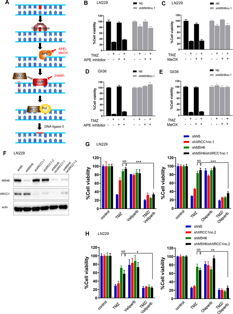
Figure 3. PARP inhibitor restoration of TMZ sensitivity is not through BER signaling blockage.
(A) A scheme of Base Excision Pathway showing PARPs, other key proteins and their inhibitors. (B, C) Non-targeting (shNS) and MSH6 knockdown (shMSH6no.1) LN229 cells were treated with TMZ (200 uM), APE inhibitor (1 uM, in B), MeOX (3 mM, in C), or TMZ combination with APE inhibitor (B) or MeOX (C). Cell viability was evaluated by Cell Titer Glo on day 6.(D,E) Same experiments as B and C using Gli36shNS and Gli36shMSH6no.1. TMZ (30 uM), APE inhibitor (1 uM, in D), MeOX (3 mM, in E) (F) LN229 cells were engineered with a non-targeting (shNS), MSH6-directed (shMSH6) or XRCC1-directed shRNA (shXRCC1–1, shXRCC1–2) lentivirus or both shMSH6 and shXRCC1. Immunoblot confirmed knockdown of MSH6 and XRCC1. Actin was used as a loading control. (G) LN229shNS, LN229shMSH6, LN229shXRCC1–1, LN229shMSH6/XRCC1–1 cells were treated with TMZ (200 uM), PARP inhibitor (veliparib, 3 uM, in E; olaparib, 1 uM, in F) or TMZ combination with PARP inhibitor. Cell viability was evaluated by Cell Titer Glo on day 6. (H) Same experiments as E and F using a second shRNA, XRCC1–2. *P<0.005, **P<0.0005, ***P<0.0001 (student t-test).

