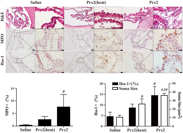
Fig. 3.

Examples of H&E stained sections myeloperoxidase (MPO; neutrophil marker) and Iba-1 (macrophage marker) immunohistochemistry at the choroid plexus one day after intraventricular saline, Prx2(heat) and Prx2 injection. The numbers of MPO and Iba-1 positive cells were quantified. Values are mean ± SD, n=6, #p<0.01 vs. saline group, ##p<0.01 vs. Prx2(heat) group. Scale bar = 50μm at low magnification, =10μm at high magnification.
