
Fig 3. Embryonic developmental delay and placental developmental defects in Daam1/2-deficient mice.
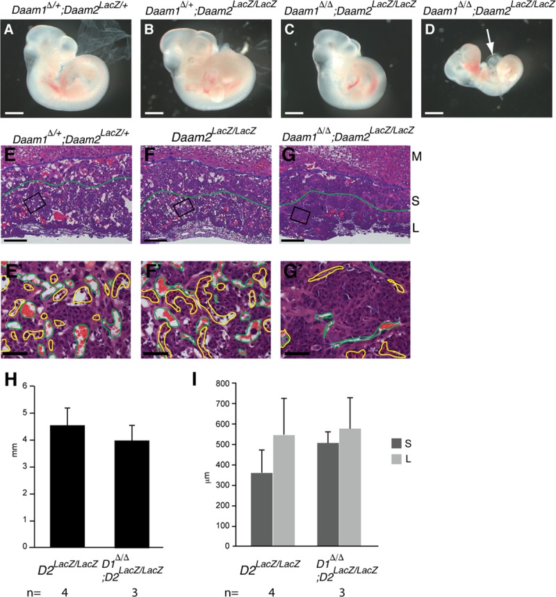
(A-C) Daam1Δ/+, Daam2LacZ/+ (A), Daam1Δ/+, Daam2 LacZ/LacZ (B), and Daam1Δ/Δ, Daam2LacZ/LacZ (C, D), embryos at E10.5. Some of Daam1Δ/Δ, Daam2 LacZ/LacZ embryos were found dead with an enlarged pericardial cavity (arrow, D). (E-G, E’-G’) H&E staining of E10.5 placenta sections of Daam1Δ/+, Daam2 LacZ/+ (E), Daam2 LacZ/LacZ (F), and Daam1Δ/Δ, Daam2 LacZ/LacZ (G). High magnification images’ (E’-G’) positions are indicated as boxes on E-G. (H) The size of the placenta of Daam2 LacZ/LacZ and Daam1Δ/Δ, Daam2 LacZ/LacZ was measured as described for Fig 2E. (I) The thickness of the spongiotrophoblast layer (S) and labyrinthine layer (L) in Daam2 LacZ/LacZ and Daam1Δ/Δ, Daam2 LacZ/LacZ, measured as described in Fig 2F. Blue lines depict the boundary between the maternal decidua (M) and spongiotrophoblast layer (S), and green lines depict the boundary between the spongiotrophoblast and labyrinthine layers (L). Maternal blood sinuses and fetal blood vessels are outlined in green and yellow, respectively (E’-G’). Scale bars = 1 mm in A-D, 200 μm in E-G, 50 μm in E’- G’.