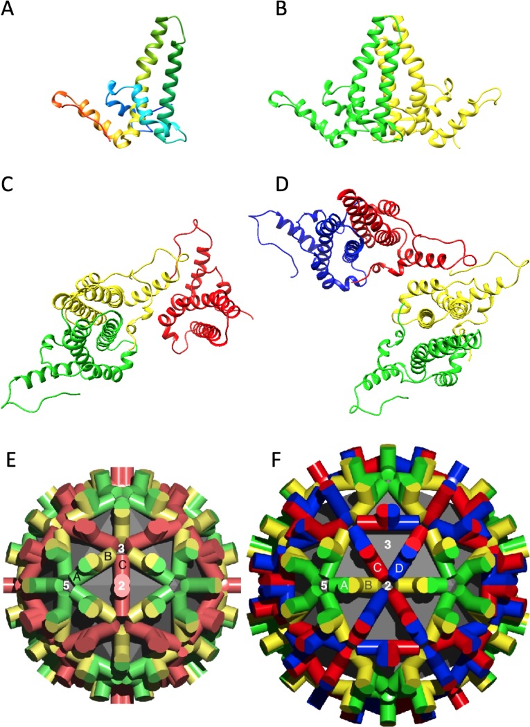
Fig 2. Conformation of core protein and its arrangement in capsids.
(A) Ribbon diagram of a single chain colored from N-terminus (blue) to C-terminus (red). Such chains do not exist in isolation. (B) An AB dimer with the A and B chains colored green and yellow, respectively. (C) The A, B, and C chains of T = 3 capsids colored green, yellow, and red, respectively. (D) The A, B, C, and D chains of T = 4 capsids colored green, yellow, red, and blue, respectively. (E, F) Lattices of the T = 3 and T = 4 capsids with the subunits labeled and colored according to the same convention. The chains in (C, D) are shown approximately as they are arranged in the corresponding lattices (E, F). The different shades in the lattices indicate quasi-equivalence.

