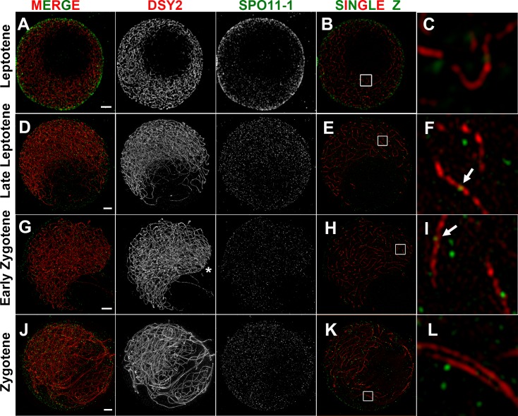
Fig 5. SPO11-1 localization revealed by super-resolution microscopy in wild-type.
Super-resolution images of SPO11-1 (green) and DSY2 (red) staining in WT meiocytes shown as projection images of nuclei (A, D, G, J), single Z sections (B, E, H, K), and magnified views of 2 μm2 regions (C, F, I, L). Scale bars represent 2 μm. Respective serial Z sections are shown in S8–S11 Figs.(A-C) A leptotene nucleus with long and intricate chromosome axes surrounding a nucleolus exhibiting most of its SPO11-1 foci around the nuclear periphery and less signal within the nucleus. (D-F) A late leptotene nucleus with an off-set nucleolus exhibiting numerous SPO11-1 foci distributed within the nucleus. No obvious pre-alignment of axes was observed (S9 Fig). Some SPO11-1 foci are located on DSY2-labeled AEs (arrow). (G-I) An early zygotene nucleus with telomere bouquet (asterisk) and pre-aligned axes (Z11-19 in S10 Fig) exhibiting numerous SPO11-1 foci. Note that the nucleolus exhibits less SPO11-1 signal. (J-L) A zygotene nucleus with progressive pairing and synapsed regions containing numerous SPO11-1 foci.

