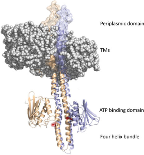
Figure 2.

EnvZ domain structure. The periplasmic domain of an EnvZ dimer protrudes above the membrane, which is shown as a space-filling model (membrane from PDB ID: 3J00, EnvZ dimer frrom PDB: 4CTI) (190). The transmembrane domains (TMs) connect to the four-helix bundle formed from a dimer of two monomers (in purple and orange); a single His243 sidechain (phosphorylation site) is highlighted in red. The ATP binding domains flank His243.
