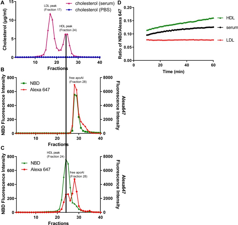
Fig. 1.
ApoA1 exchange indicator exclusively exchanges into HDL fraction in human serum. 100 µg apoA1 exchange indicator was incubated with 100 µL PBS (blue symbols) or normal human serum (pink symbols) in a total volume of 300 µL at 37°C for 1 h, and 100 µL of the product was size fractioned by FPLC using a Superose 6 (10/300 GL) column and 0.5 ml fractions were collected. The cholesterol concentration was measured in each fraction (A), with the LDL-C peak at fraction 17 and HDL-C peak at fraction 24. The fluorescent intensities of NBD (green symbols) and Alexa647 (red symbols) were measured in each fraction for the apoA1 exchange indicator incubated with PBS only (B), or incubated with human serum (C). D. 5 µg apoA1 exchange indicator was incubated with serum (black symbols), human LDL (5 µg LDL-C, red symbols), or human HDL (2.5 µg HDL-C, green symbols) at 37°C in a 96 well dish the NBD/Alexa647 fluorescence ratio determined at 1 min intervals over 1 h (excluding the nonlinear first 10 min), demonstrating the method used to calculate AER.

