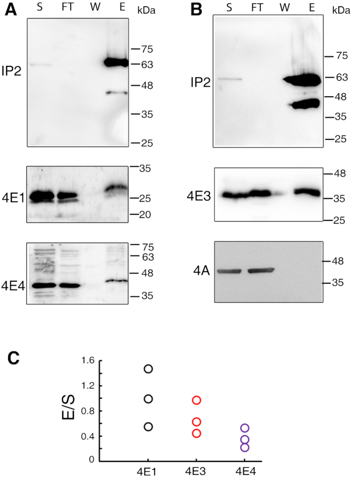
Figure 1.

Association of Leish4E-IP2 with different LeishIF4Es. The association between Leish4E-IP2 and LeishIF4E-1 (4E1), LeishIF4E-4 (4E4) (A) and LeishIF4E-3 (4E3) (B) were monitored by pull-down experiments of mid-log L. amazonensis promastigotes over-expressing SBP-tagged LeishIF4E-IP2. The cells were lysed and affinity-purified over streptavidin-Sepharose beads. The beads were washed and further eluted with biotin. Aliquots from the soluble extract (S, 5%), the flow-through fraction (FT, 5%), the final wash (W, 50%) and eluted proteins (E, 50%) were separated by 10% SDS–PAGE and subjected to western blot analysis using specific antibodies. The antibodies used were raised against LeishIF4E-1, LeishIF4E-4 (A), LeishIF4E-3 and LeishIF4A (as a negative control) (B) along with anti-SBP antibodies that were used to highlight the SBP-tagged Leish4E-IP2. (C) Representation of the densitometry analysis of each quantity of LeishIF4E co-eluted by Leish4E-IP2.
