Figure 3.

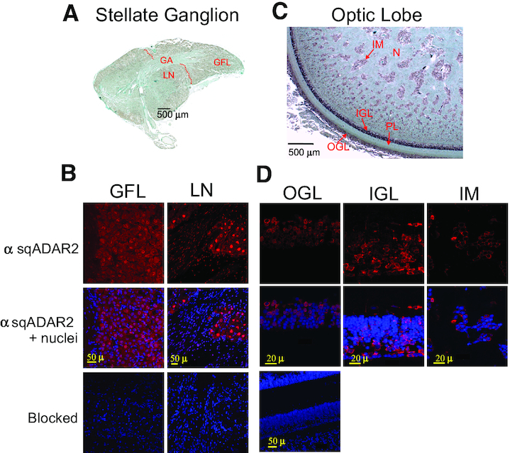
sqADAR2 is expressed in the cytoplasm of squid somata. (A) Sagittal section from the SG of D. pealeii. The GFL within the SG, large neurons (LN) outside the GFL and the squid GA are shown. A dashed red line delineates the border between the GFL and the rest of the SG. (B) Immunostaining of sqADAR2 proteins in both squid GFL and LN within the SG. Cells were stained with α-sqADAR2 primary antibody, while DAPI and To-Pro-3 were used for nuclear visualization. Blocked controls are also shown. (C) Sagittal section from the OL of D. pealeii. The structures comprising the OL including the outer granular layer (OGL), inner granular layer (IGL), plexiform layer, islands of the medulla (IM) and nuclei are shown. (D) Immunostaining of sqADAR2 proteins in neuronal cells within the squid OL. Cells were stained with α-sqADAR2 primary antibody, while DAPI and To-Pro-3 were used for nuclear visualization. Blocked controls are also shown. Cells were imaged using a Zeiss LSM 510 laser scanning confocal microscope.
