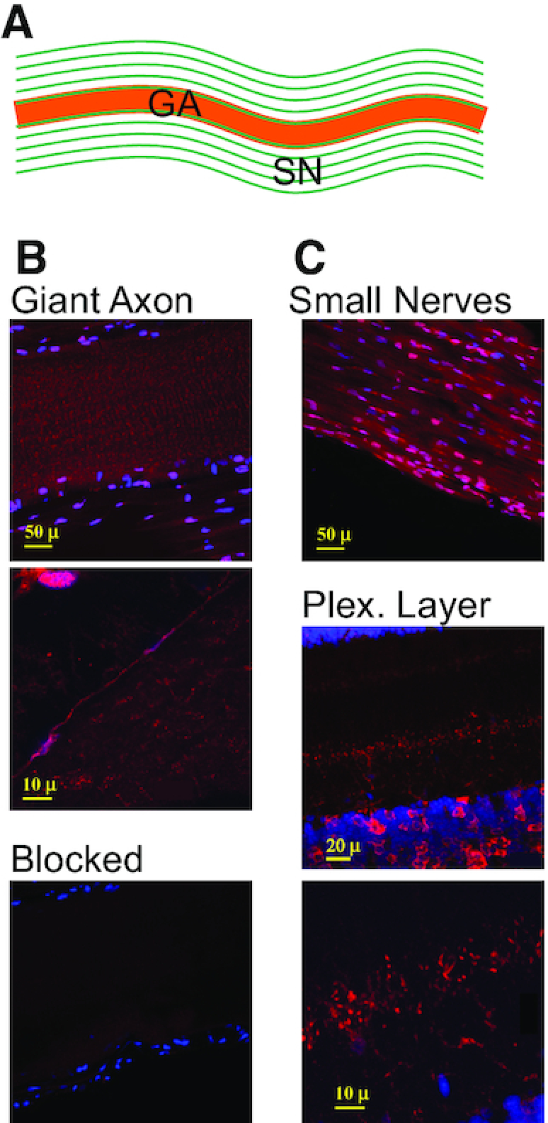
Figure 4.

sqADAR2 is expressed in squid axons. (A) Diagram of the squid GA with its surrounding small nerve axons (SN) running in parallel. (B) Immunostaining of sqADAR2 proteins in squid GA (i). Higher magnification of same area is shown (ii). (C) Immunostaining of sqADAR2 proteins in the SN (i) and plexiform layer of the OL (ii). Higher magnification of same area or plexiform layer is shown (iii). Cells were stained with α-sqADAR2 primary antibody, while DAPI and To-Pro-3 were used for nuclear visualization. Blocked control is also shown (B, iii). Cells were imaged using a Zeiss LSM 510 laser scanning confocal microscope.
