
Figure 4.
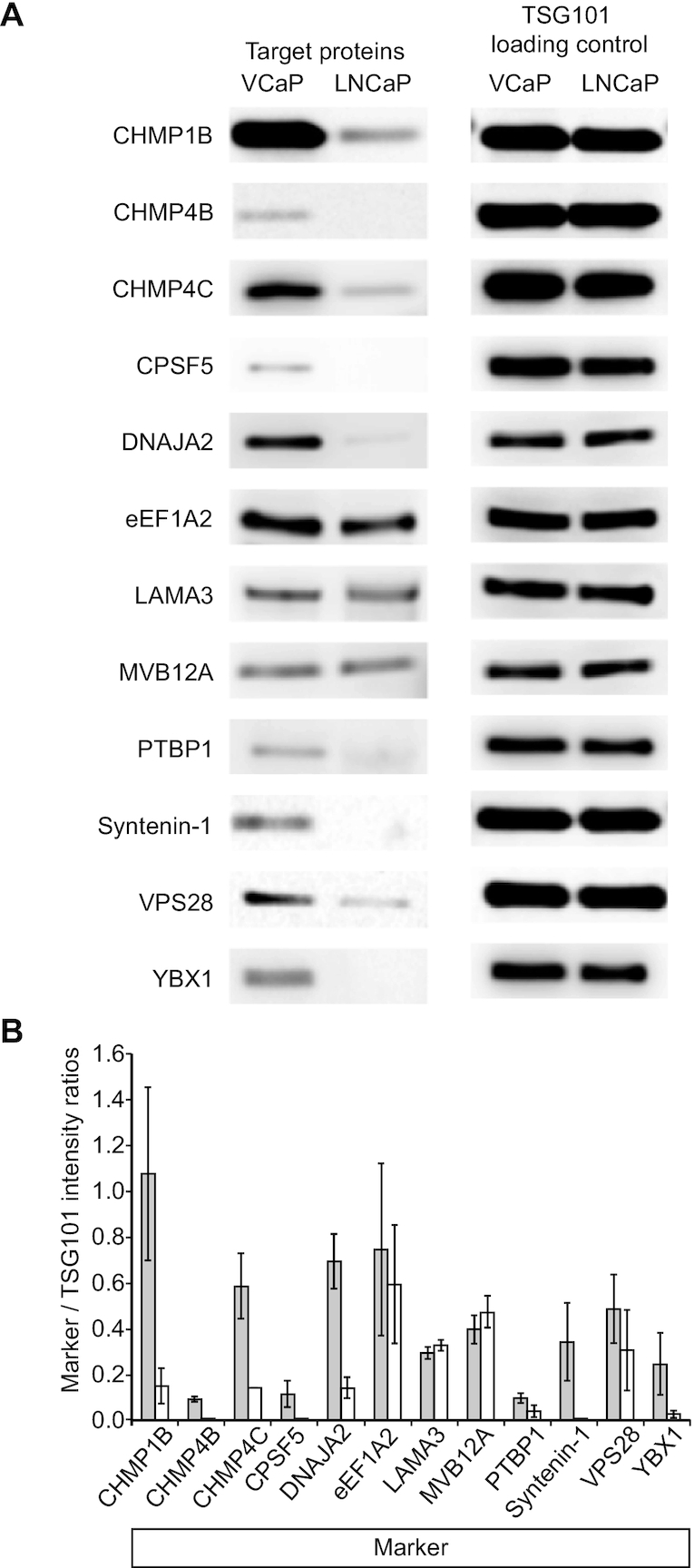
Differential detection of identified proteins in VCaP vs LNCaP exosomes by western blotting. (A) Of the 12 proteins tested (CHMP1B, CHMP4B, CHMP4C, CPSF5, DNAJA2, PTBP1, Syntenin-1 and YBX1 all exhibit higher expression in exosomes from VCaP cells (VCaP) compared to exosomes from LNCaP cells (LNCaP) as quantified by the intensity ratios of targets to the respective TSG101 loading and transfer controls. Only eEF1A2, LAMA3 and MVB12A showed similar expression levels in VCaP and LNCaP exosomes. (B) Relative quantification of intensities from several western blot experiments (VCaP exosomes: dark bars; LNCaP exosomes: light bars) in relation to the respective TSG101 intensities. CHMP1B: n = 4; CHMP4B: n = 2; CHMP4C: n = 2; CPSF5: n = 2; DNAJA2: n = 3; EEF1A2: n = 2; LAMA3: n = 2; MVB12A: n = 2; PTBP1: n = 2; Syntenin-1: n = 2; VPS28: n = 4; YBX1: n = 2. Error bars: mean ± S.D. All proteins verified by western blotting are shown in italic in Figure 3C. We tested all proteins listed in Table 1 and Figure 3C, respectively, but obtained ambiguous results due to insufficient antibody specificity (data are not shown).