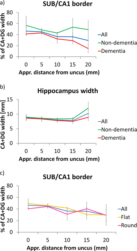
Figure 12.
SUB/CA1 border in the hippocampal body along the longitudinal axis and potential confounds. a. SUB/CA1 border relative to the CA+DG width in the whole group and separately in the non-dementia and dementia subjects. b. Overall hippocampus width in the whole group and separately in the non-dementia and dementia subjects. c. SUB/CA1 border relative to the CA+DG width in the whole group and separately in the flat- and round-hippocampi specimen.
CA=cornu ammonis; DG=dentate gyrus; SUB=subiculum

