
Figure 3: PDK1 inhibition regulates proplatelet formation and MARCKs expression.
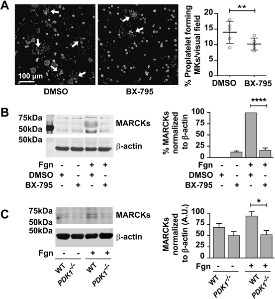
A. CD34+ megakaryocytes are treated with BX795 (1μM) from day 11 – 13. Cells are collected on Day 13 and adheared to fibrinogen coated plates and incubated over night at 370C. The cells are then treated with phalloidin dye and observed for proplatelet formation using confocal microscopy. The number of proplatelet forming cells were counted and graphs are plotted. B. CD34+ megakaryocytes are treated with BX795 (1μM) from day 11 – 13. Cells are collected on Day 13 and adheared to fibrinogen for 2 hours. Proteins were separated by using SDS-PAGE and probed from MARCKs protein. The immunoblot is representative of three independent experiments (****P < 0.05). C. Day 5 bone marrow derived mouse megakaryocytes from wild type or platelet specific PDK1 knockout mice were analyzed for expression of MARCKs proteins under adherent or non-adherent conditions on fibrinogen using SDS-PAGE. The western blots shown are representative of three independent experiments. (*P < 0.05).