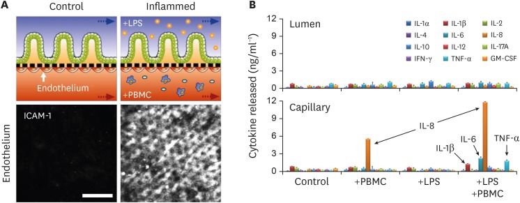
Figure 2. Demonstration of the pathophysiological immune responses in the gut-on-a-chip challenged to LPS and PBMCs. (A) The schematics in the upper layer show the experimental design of the healthy (control) versus stimulated microenvironment (inflamed) of the intestinal tissue interface composed of the intestinal villus epithelium and vascular endothelium. In this setup, inflammatory responses are induced by introducing LPS and PBMC at the upper and the lower microchannels, respectively, for 48 h. Confocal micrographs confirm the strong activation of ICAM-1 on the apical surface of the capillary endothelium when the EMI axis is perturbed and inflamed. Bar, 50 μm.(B) The basolateral secretion of proinflammatory cytokines (IL-1β, IL-6, IL-8, and TNF-α) illustrates the mucosal inflammation in response to the co-stimulation of LPS or E. coli cells and PBMCs. It is noted that a directional release of the proinflammatory cytokines demonstrates possible immune cell recruitment from the lamina propria in vitro. Data were reorganized from the references (12).
ICAM-1, intercellular adhesion molecule 1.

