Fig. 3.
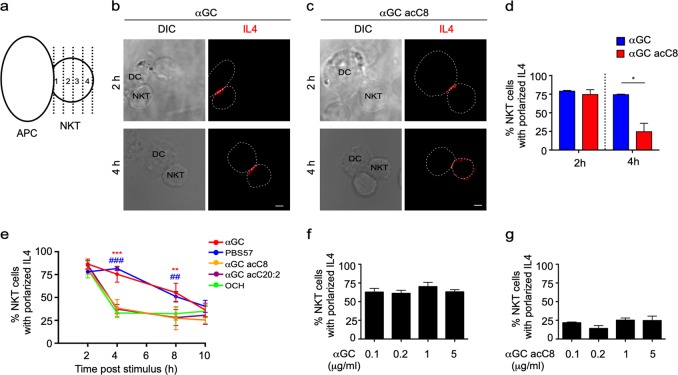
α-Galactosylceramide (αGC), not T-helper type 2 (Th2) lipid antigens, maintains the polarized secretion of IL4 in invariant natural killer T (iNKT) cells. a Method to quantify the polarized secretion of IL4. IL4 in area 1 is defined as polarized secretion. b, c Secretory sites of IL4 in iNKT cells activated by distinct antigen variant-pulsed splenic dendritic cells (DCs) at the indicated time points. Dotted lines indicate cell boundaries. Scale bars, 2 μm. Data are representative of three independent experiments and more than 70 cells per group. d Frequency of iNKT cells with polarized IL4 secretion. Data are presented as the mean ± SEM of three independent experiments. e Frequency of iNKT cells with polarized IL4 secretion after activation by distinct antigen variant-pulsed RBL.CD1d cells at the indicated time points. Data are presented as the mean ± SEM of three independent experiments. * indicates a significant difference between αGC and Th2 lipids, and # indicates a significant difference between PBS57 and Th2 lipids. f, g Frequency of polarized IL4 secretion at the IS induced by the indicated concentrations of αGC (f) and αGC acC8 (g) 4 h after activation. Data are presented as the mean ± SEM of three independent experiments. Statistical analysis was performed using Student’s t -test, two-way analysis of variance (ANOVA) or one-way ANOVA with the Tukey’s post test. *P < 0.05; ** or ##P < 0.01; and *** or ###P < 0.001

