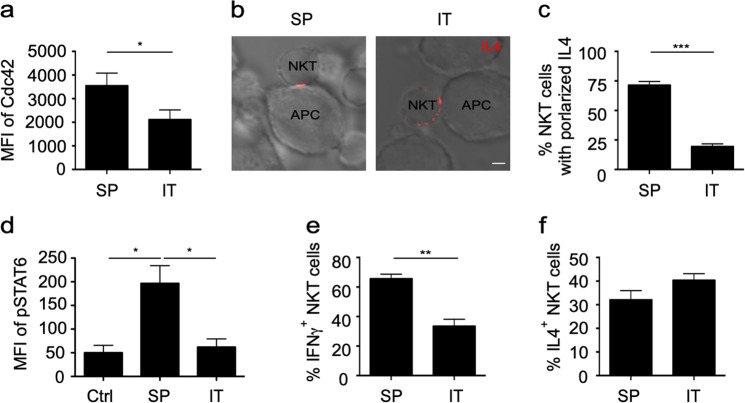
Fig. 7.
Impaired interleukin-4 (IL4) polarization and invariant natural killer T (iNKT) cell-dendritic cell (DC) crosstalk in tumors. a Expression of Cdc42 in intratumoral (IT) and splenic (SP) iNKT cells from MC38 tumor-bearing mice. Data are presented as the mean ± SEM of more than nine mice per group. b, c IL4 secretory sites (b) and the frequency of (c) intratumoral and splenic iNKT cells with polarized secretion after activation by αGC-pulsed RBL.CD1d for 4 h. Scale bars, 2 μm. Data are representative of three independent experiments (more than 60 cells per group, b) or are presented as the mean ± SEM of three independent experiments (c). d–f Phosphorylated signal transducer and activator of transcription 6 (STAT6) in splenic DCs and intratumoral MHC II+ CD24+ F4/80− CD11c+ DCs (d) and the percentages of IFNγ+ (e) and IL4+ (f) iNKT cells in the tumor and spleen of MC38 tumor-bearing mice 8 h after injection with αGC. Data are presented as the mean ± SEM of more than nine mice. Statistical analysis was performed using the Mann–Whitney U test or Student’s t test. *P < 0.05; **P < 0.01; and ***P < 0.001

