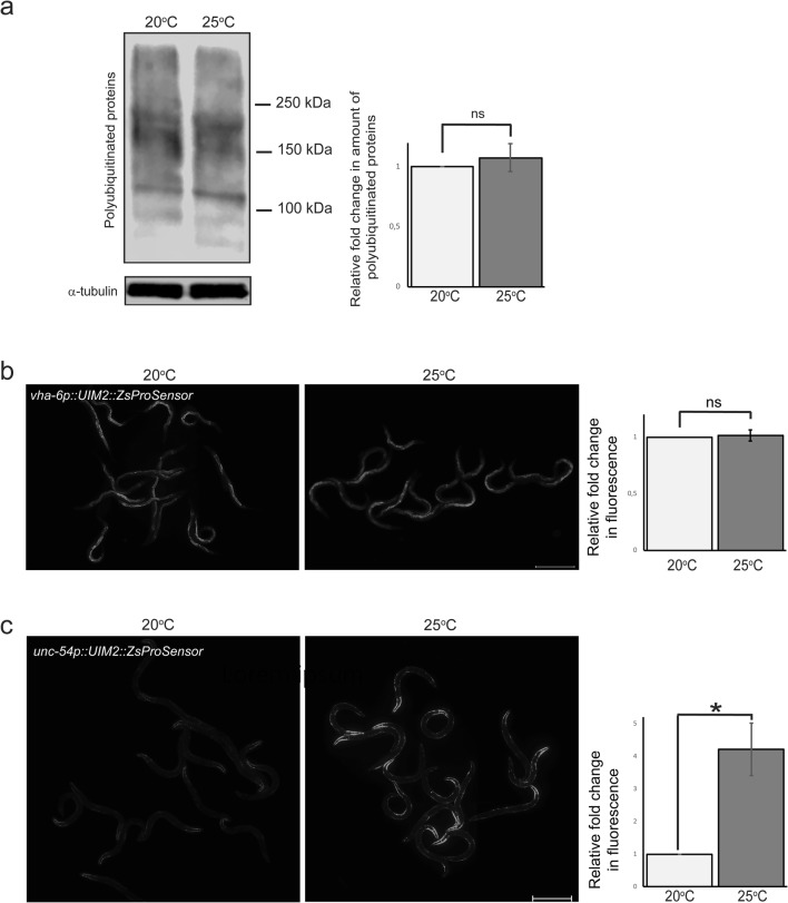
Fig. 4.
Polyubiquitinated proteins accumulate in body wall muscle at 25 °C. a Western blot against polyubiquitinated proteins (left) and quantification (right) of whole animal lysates of animals exposed to 20 °C or 25 °C. Anti-alpha-tubulin antibody was used as a normalisation control. Graph shows the average fold change compared with 20 °C (set to 1) and is the mean of 8 independent experiments. b No change is detectable in fluorescence intensity of the intestinal polyubiquitin reporter at 25 °C but c muscle reporter fluorescence increases approximately fourfold. Graphs (on right) show average fold change in fluorescence intensity compared with 20 °C (set to 1) and are the mean of a minimum of three independent experiments (n = 119 animals per temperature). UIM2 = ubiquitin-interacting motifs; ZsProSensor = ZsGreen::MODC transgene; MODC = C-terminal mouse ornithine decarboxylase. Error bar, SEM; ns, not significant; *p value, < 0.05. Scale bar, 500 μm

