Figure 1.
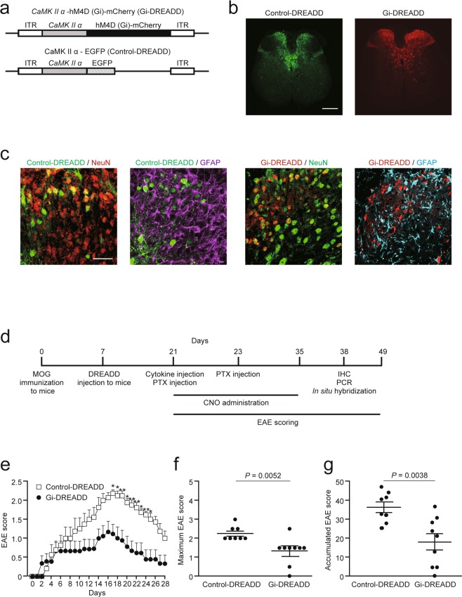
CNO-gated DREADD treatment suppressed EAE severity. (a) Schematic illustration of pAAV-CaMKIIα-hM4Di (Gi)-mCherry (Gi-DREADD) and pAAV-CaMKIIα-EGFP (control-DREADD). The AAV9 vector contains Gi- or control-DREADD under the CaMKIIα promoter. (b) Distribution of control- and Gi-DREADD at spinal level Th 8 in mice 14 days after injection of DREADD-carrying-AAV9. Scale bars: 150 μm. (c) Higher magnification images showing DREADD colocalization with NeuN or GFAP in the dorsal column of the Th 8 level spinal cord. NeuN (red) and GFAP (magenta) in control-DREADD (green) experiment. NeuN (green) and GFAP (cyan) in Gi-DREADD (red) experiment. Scale bar: 50 μm. (d) Experimental schema. Targeted EAE was induced with MOG35–55 peptide and local injection of TNFα and IFNγ. Seven days after MOG immunization, DREADD-carrying-AAV9 was injected at spinal level Th 9. EAE scores were assessed from day 0 to 28 after cytokine injection. (e) EAE scores throughout the 28 days (control: n = 8, Gi: n = 9; two-way ANOVA followed by the Sidak test). (f) Maximum and (g) accumulated EAE scores (control: n = 8, Gi: n = 9; Student’s t-test). Data are presented as mean ± sem.

