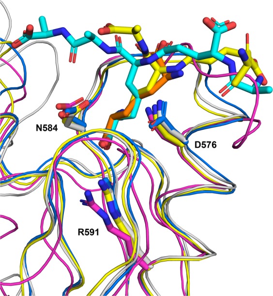
Figure 4.

Superimposition of the PorE OmpA_C-like domain structure with other representative OmpA family proteins (same as in Fig. 3). Structures of the proteins are displayed as a ribbon diagram with worm format; the peptidoglycan (PG) fragments as well as the three conserved residues whose side chain is involved in hydrogen bonds with the PG fragment are displayed in stick format, with nitrogen and oxygen atoms displayed in blue and red, respectively. The protein chains and their bound PG fragments are respectively shown in white and grey (PorE OmpA_C-like domain), yellow and orange (A. baumannii PAL, 4G4V), and blue and cyan (A. baumannii OmpA, 3TD5). V. alginolyticus PomB (3WPW), that contains no PG fragment, is shown in magenta. For clarity purpose, only the PorE OmpA_C-like domain residues are labelled, and the orientation is rotated 90° from Figs. 1 and 2 along the vertical axis. The figure was prepared using PyMOL (version 1.20, https://pymol.org).
