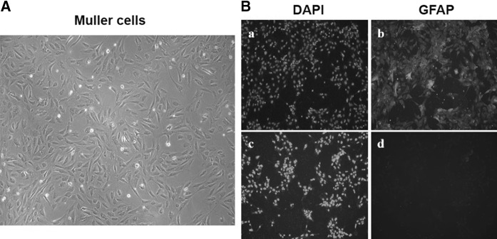
Fig. 1.
Results of determining the purity of Müller cells. Morphology of Müller cells a in good condition was photographed and then the immunofluorescence b was performed to detect the purity of Müller cells: a the DAPI staining for nucleus of Müller cells; (b, d) the green fluorescence of GFAP in Müller cells; (c) the DAPI staining for nucleus of Müller cells with the secondary antibodies conjugated-FITC; d the Müller cells just with the secondary antibodies conjugated-FITC as control

