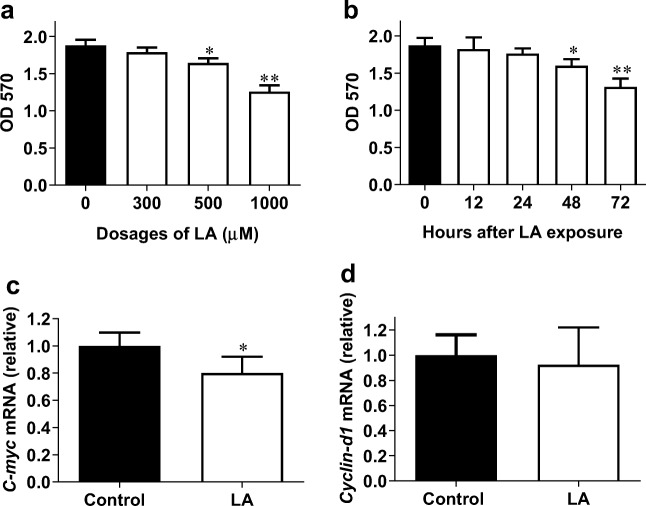
Fig. 1.
LA decreased viable HUVECs. a HUVECs were treated with LA at the indicated dosages for 48 h. MTT assay was performed to evaluate viable cells. **P < 0.01 and *P < 0.05 vs. untreated (0 μM) control cells, n = 3/group. b HUVECs were treated with LA (500 μM) for the indicated durations. MTT assay was performed to evaluate viable cells. **P < 0.01 and *P < 0.05 vs. 0 h control cells, n = 3/group. c HUVECs were incubated with LA (500 μM) for 48 h. Cells were collected for analysis of C-myc mRNA expression using real-time PCR. *P < 0.05 vs. untreated control cells, n = 4/group. d HUVECs were incubated with LA (500 μM) for 48 h. Cells were collected for analysis of Cyclin-D1 mRNA expression using real-time PCR. *P < 0.05 vs. untreated control cells, n = 3/group. LA, α-lipoic acid

