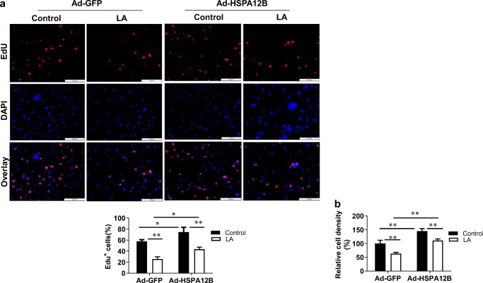
Fig. 8.
Overexpression of HSPA12B attenuated the LA-induced inhibition of viable HUVECs. HUVECs were infected with HSPA12B adenovirus (Ad-HSPA12B) or empty adenovirus (Ad-GFP). Twenty-four hours later, cells were incubated with LA (500 μM) for 48 h. Untreated cells served as controls. HUVEC proliferation alterations were assessed using cell counts and EdU labeling, then photographed with a fluorescence microscope at magnification of × 100. Scale bar represents 200 μm. **P < 0.01 vs. untreated control cells, n = 3/group. LA, α-lipoic acid. **P < 0.01 or *P < 0.05, n = 3/group. LA, α-lipoic acid. Scale bar = 100 μM

