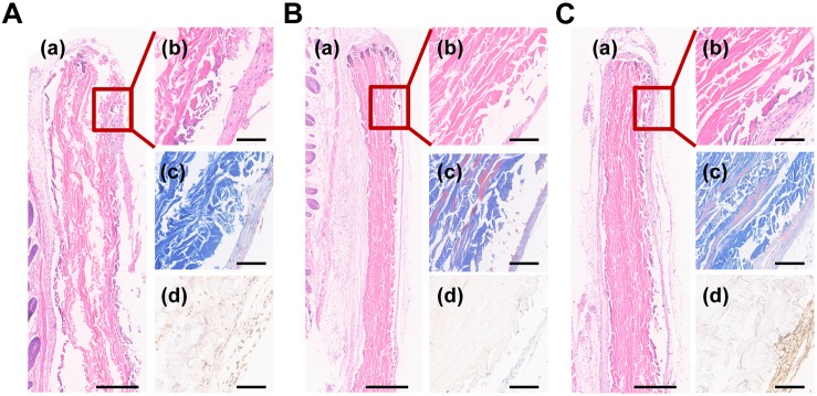
FIGURE 7.
Histological images of (A) ACL and (B) biomimetic cementum without PDLCs and (C) biomimetic cementum seeded with PDLCs. (a,b) indicated the HE staining, (c) indicated Masson trichrome staining, and (d) indicated immunohistochemical staining of CEMP1. Scale bar in a of A–C: 500 μm. Scale bar in b–d of A–C: 100 μm.

